Sympathetic connections of the pterygopalatine and superior cervical ganglia
About this plate
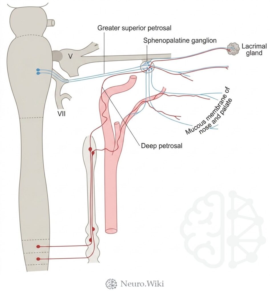
Sympathetic connections of the pterygopalatine and superior cervical ganglia.
The pterygopalatine fossa is a small pyramidal space bounded anteriorly by the maxilla, posteriorly by the sphenoid pterygoid process, and medially by the palatine bone, entered laterally through the pterygomaxillary fissure. It houses the pterygopalatine ganglion and the maxillary nerve, which gives posterior, middle, and anterior superior alveolar branches to the upper teeth. The vidian nerve, formed by the greater superficial petrosal from the facial nerve and the deep petrosal carrying sympathetic fibres from the superior cervical ganglion, joins the ganglion; postganglionic fibres reach the lacrimal gland and the mucous membrane of the nose and palate.
Structures labelled on this plate
- Greater superior petrosal
- Sphenopalatine ganglion
- Lacrimal gland
- Vii
- Mucous membrane of nose and palate
- Deep petrosal
Source: After Gray's Anatomy (1918). Public domain (Gray's Anatomy, 1918), modernized rendering.
Rendering © 2026 Neuro.Wiki — recolouring, relabelling and three-dimensional modelling. The underlying historical figure is public domain and remains so. Free to view and to cite with a link; not licensed for redistribution, dataset compilation, or AI model training.