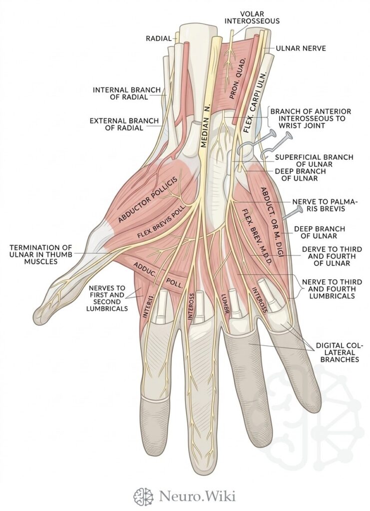
Deep palmar nerves of the left hand, palmar view
About this plate
Deep palmar nerves of the left hand, palmar view.
The palmar aspect exposes the flexor compartment, covered by the palmar aponeurosis and, at the wrist, by the transverse carpal ligament roofing the carpal tunnel. Through it pass the median nerve and the tendons of flexor digitorum superficialis and profundus in a common synovial sheath, with the separate sheath of flexor pollicis longus. The thenar eminence carries abductor pollicis brevis, opponens pollicis and flexor pollicis brevis, with adductor pollicis deeper. The hypothenar mound contains palmaris brevis and abductor digiti minimi. Lumbricals and the deep ulnar branch complete intrinsic function; radial and ulnar arteries form the palmar arches and digital vessels.
Structures labelled on this plate
- Radial
- Volar interosseous
- Ulnar nerve
- Internal branch of radial
- External branch of radial
- Median nerve
- Pron. quad
- Flexor carpi uln
- Branch of anterior interosseous to wrist joint
- Superficial branch of ulnar
- Deep branch of ulnar
- Abductor pollicis
- Flexor brevis poll
- Nerve to palma-ris brevis
- Termination of ulnar in thumb muscles
- Abduc. poll
- Nerve to third and fourth of ulnar
- Flexor brevis muscle d
- Nerve to first and second lumbricals
- Nerve ii
- Inteross
- Lumbr
- Nerve to third and fourth lumbricals
- Digital collateral branches
Source: After Gray's Anatomy (1918). Public domain (Gray's Anatomy, 1918), modernized rendering.
Rendering © 2026 Neuro.Wiki — recolouring, relabelling and three-dimensional modelling. The underlying historical figure is public domain and remains so. Free to view and to cite with a link; not licensed for redistribution, dataset compilation, or AI model training.