Pelvic surface of the sacrum
About this plate
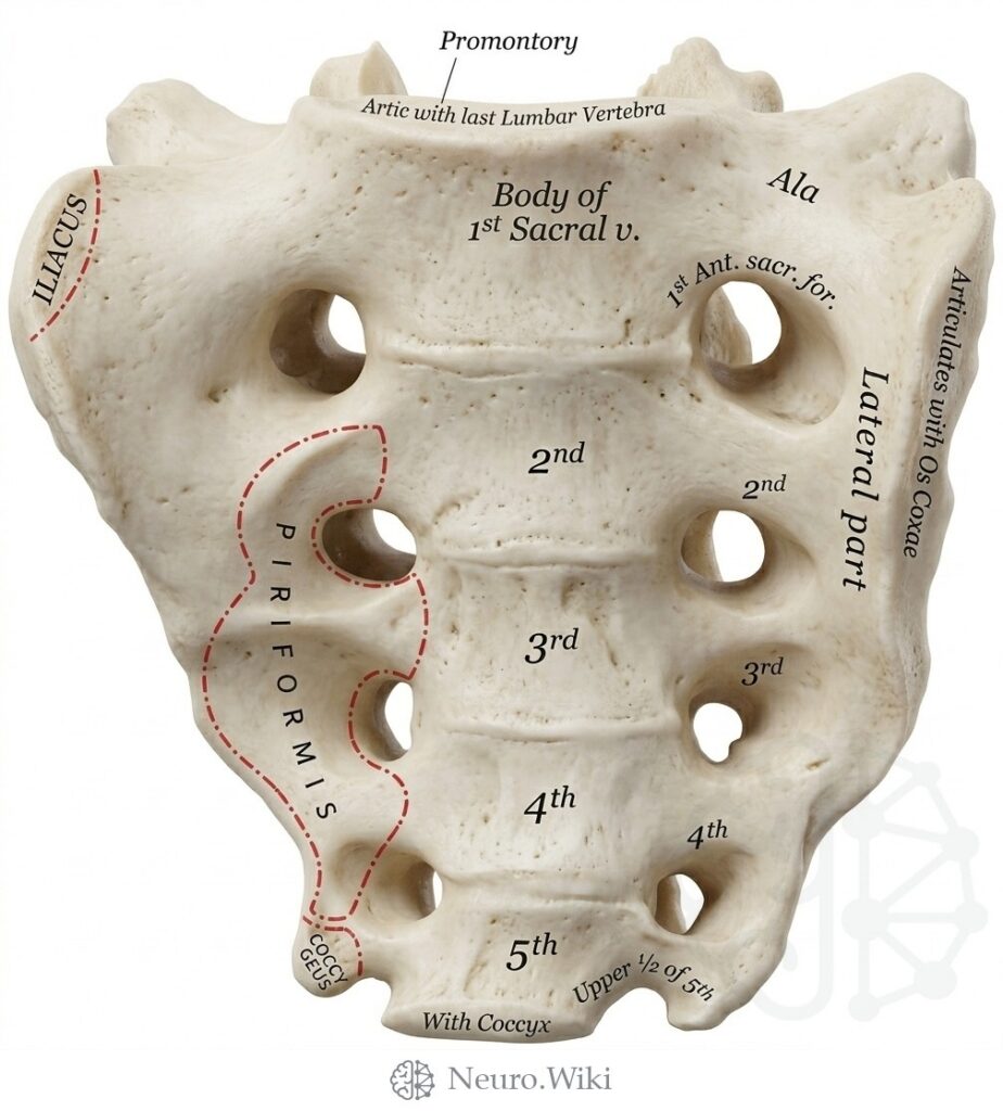
Pelvic surface of the sacrum.
The sacrum is a large triangular bone formed by fusion of five sacral vertebrae, wedged between the two hip bones and articulating superiorly with the fifth lumbar vertebra at the prominent sacral promontory. Its concave pelvic surface shows four transverse ridges marking the fused vertebral bodies, with paired anterior sacral foramina transmitting the ventral rami of the upper sacral nerves. Laterally, the alae and auricular surfaces articulate with the ilia. The piriformis arises from its pelvic aspect. Below, the small triangular coccyx, usually four rudimentary vertebrae, articulates with the sacral apex and gives attachment to coccygeus and levator ani.
Structures labelled on this plate
- Promontory
- Artic with last Lumbar Vertebra
- Iliacus
- Body of 1st Sacral vein
- Ala
- 1st anterior sacr. foramen
- Articulates with Os Coxæ
- Lateral part
- Piriformis
- Coccygeus
- Upper 1/2 of 5th
- With Coccyx
Source: After Gray's Anatomy (1918). Public domain (Gray's Anatomy, 1918), modernized rendering.
Rendering © 2026 Neuro.Wiki — recolouring, relabelling and three-dimensional modelling. The underlying historical figure is public domain and remains so. Free to view and to cite with a link; not licensed for redistribution, dataset compilation, or AI model training.