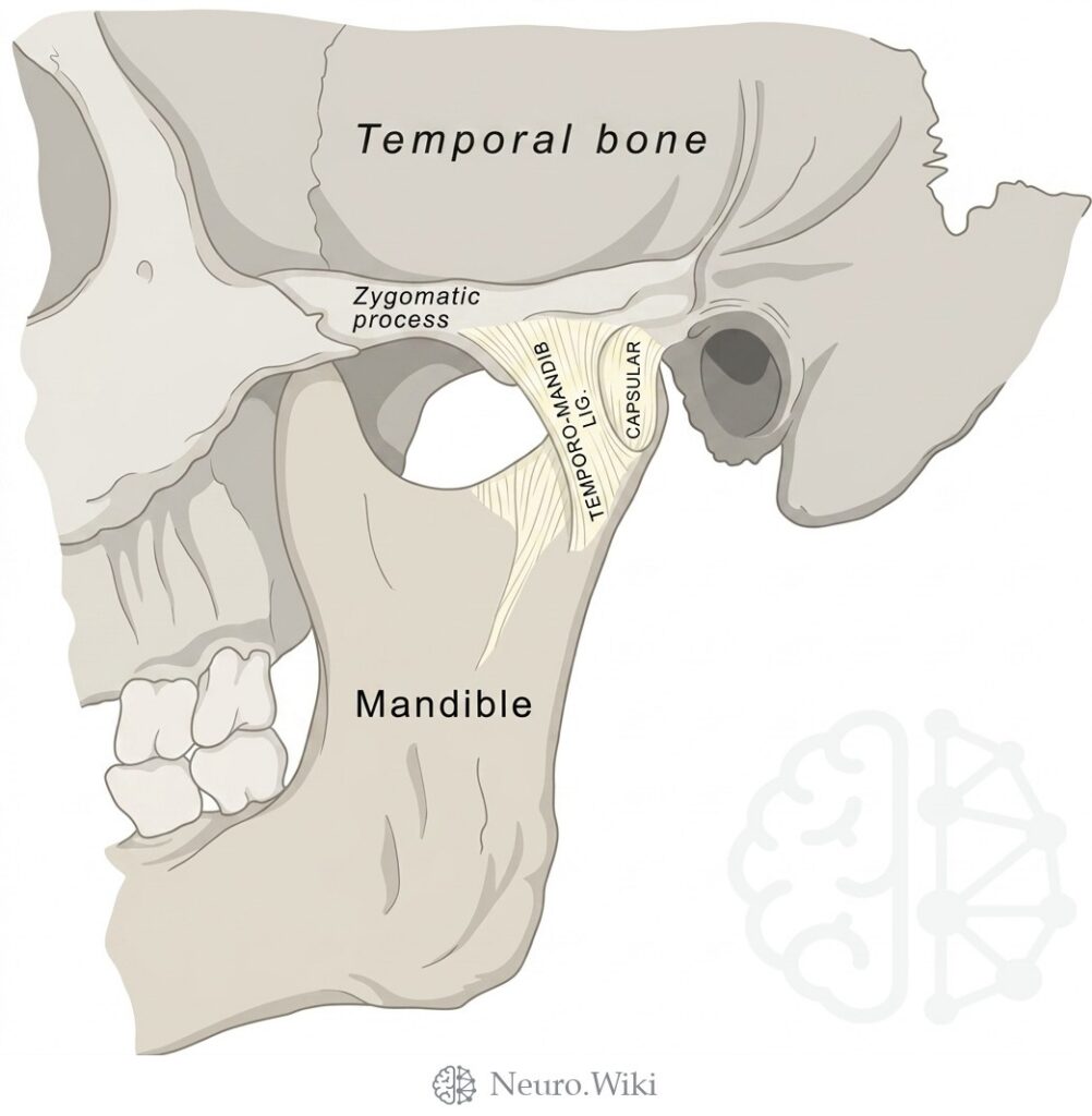
Temporomandibular joint, lateral view
About this plate
Temporomandibular joint, lateral view.
The mandible is the U-shaped bone of the lower jaw, the only mobile bone of the skull. In lateral profile it presents a horizontal body and a vertical ramus meeting at the angle. The body carries the alveolar border for the lower dentition and, anteriorly, the mental protuberance and paired mental foramina, which transmit the mental neurovascular bundles. The oblique line sweeps up from the body onto the ramus, giving attachment to depressors of the lower lip, including depressor labii inferioris, depressor anguli oris, and platysma; buccinator arises further back along the alveolar margin. Superiorly the ramus divides into an anterior coronoid process, receiving temporalis, and a posterior condylar process, whose head and neck articulate at the temporomandibular joint with the mandibular fossa of the temporal bone beneath its zygomatic process. Between them lies the mandibular notch. The lateral surface of the ramus receives masseter, and the temporomandibular ligament reinforces the joint capsule laterally.
Structures labelled on this plate
- Temporal bone
- Zygomatic process
- Temporo-mandib ligament
- Capsular
- Mandible
Source: After Gray's Anatomy (1918). Public domain (Gray's Anatomy, 1918), modernized rendering.