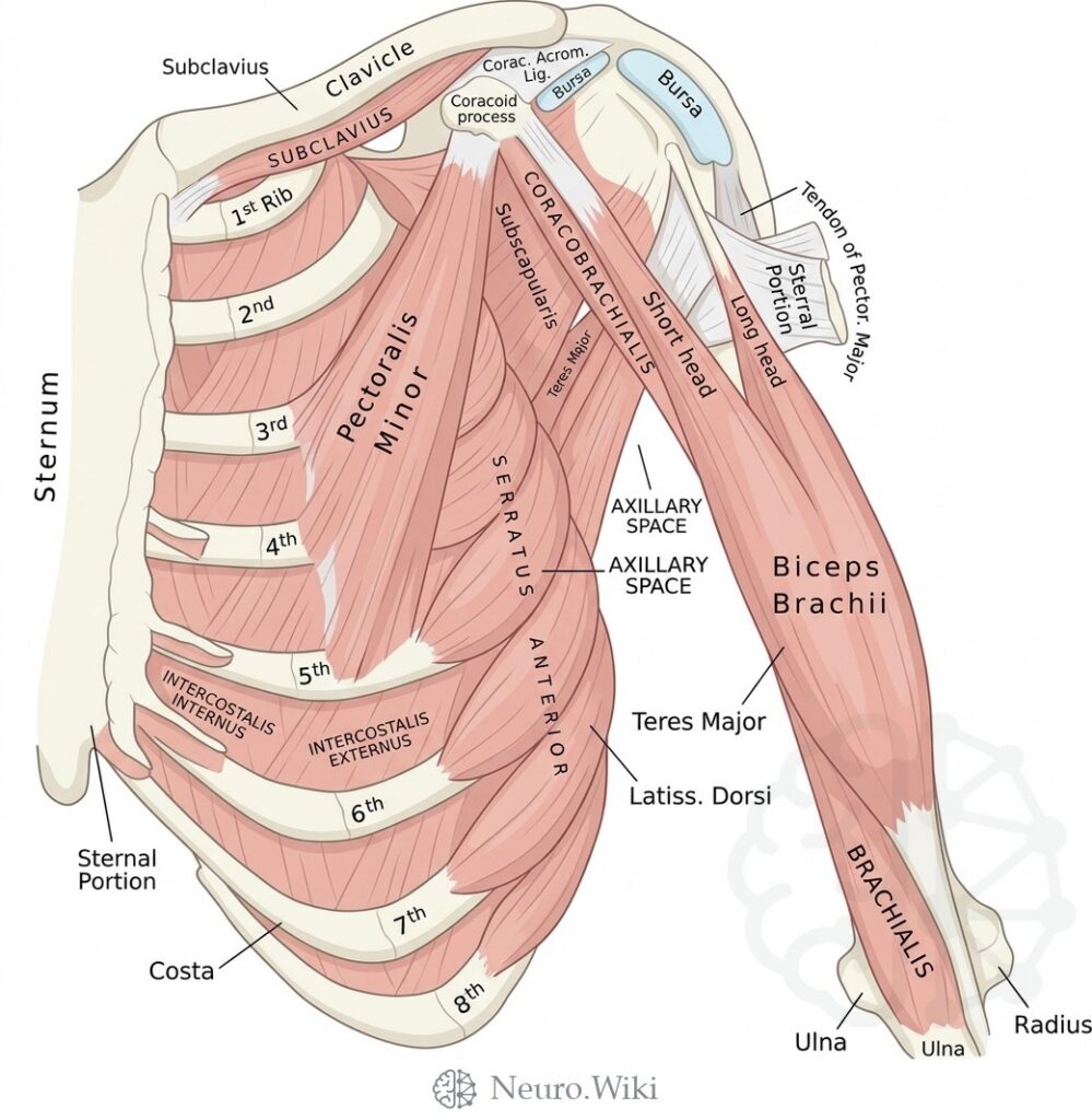
Anterior view of deep chest and arm muscles with axillary boundaries
About this plate
Anterior view of deep chest and arm muscles with axillary boundaries.
The anterior thoracic wall is framed by the sternum and the paired costal cage curving laterally, with the intercostal spaces occupied by the external and internal intercostal muscles. Superficial to this bony scaffold lie the pectoral muscles; deep to pectoralis major sit pectoralis minor, arising from the anterior ribs to reach the coracoid process, and subclavius, bridging the first rib to the clavicle. Serratus anterior sweeps across the lateral wall onto the scapula. The axillary space opens between the pectoral group and subscapularis, teres major and latissimus dorsi, transmitting coracobrachialis and both heads of biceps brachii toward brachialis.
Structures labelled on this plate
- Subclavius
- Clavicle
- Corac. Acrom. ligament
- Bursa
- Coracoid process
- 1st Rib
- Pectoralis Minor
- Subscapularis
- Coracobrachialis
- Short head
- Longus head
- Sternal Portion
- Tendon of Pector. Major
- Teres Major
- Axillary space
- Sternum
- Serratus anterior
- Biceps Brachii
- Intercostalis internus
- Intercostalis externus
- Latiss. Dorsi
- Costa
- Brachialis
- Ulna
- Radius
Source: After Gray's Anatomy (1918). Public domain (Gray's Anatomy, 1918), modernized rendering.
Rendering © 2026 Neuro.Wiki — recolouring, relabelling and three-dimensional modelling. The underlying historical figure is public domain and remains so. Free to view and to cite with a link; not licensed for redistribution, dataset compilation, or AI model training.