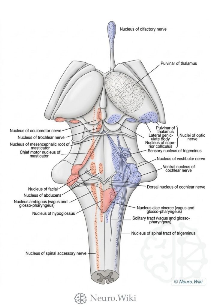
Scheme showing the Relative Size and Position of the Nuclei of Origin (Red) of the Motor and the Nuclei of Termination (Blue) of the Sensory Cranial Nerves
About this plate
Scheme showing the Relative Size and Position of the Nuclei of Origin (Red) of the Motor and the Nuclei of Termination (Blue) of the Sensory Cranial Nerves.
Viewed from behind, the brainstem exposes dorsal midbrain, pons and medulla once the cerebellum is removed. The corpora quadrigemina—paired superior and inferior colliculi—cap the midbrain, with the pineal body and habenular commissure above and the pulvinar and medial geniculate bodies laterally. The trochlear nerves emerge just below the inferior colliculi at the frenulum of the anterior medullary velum. The rhomboid fossa forms the fourth-ventricular floor, showing the medial eminence, facial colliculus and locus caeruleus between the cerebellar peduncles. Caudally, the gracile and cuneate fasciculi end in the clava and cuneate tubercle at the obex.
Structures labelled on this plate
- Nucleus of olfactory nerve
- Pulvinar of thalamus
- Nucleus of oculomotor nerve
- Nucleus of trochlear nerve
- Nucleus of mesencephalic root of masticator
- Chief motor nucleus of masticator
- Lateral genic- ulate body
- Nuclei of optic nerve
- Nucleus of supe- rior colliculus
- Sensory nucleus of trigeminus
- Nucleus of vestibular nerve
- Ventral nucleus of cochlear nerve
- Nucleus of facial
- Nucleus of abducens
- Dorsal nucleus of cochlear nerve
- Nucleus ambiguus (vagus and glosso-pharyngeus)
- Nucleus alae cinereae (vagus and glosso-pharyngeus)
- Nucleus of hypoglossus
- Solitary tract (vagus and glosso- pharyngeus)
- Nucleus of spinal tract of trigeminus
- Nucleus of spinal accessory nerve
Source: After Morris's Human Anatomy (1918). Public domain (Morris's Human Anatomy, 1918), modernized rendering.
Rendering © 2026 Neuro.Wiki — recolouring, relabelling and three-dimensional modelling. The underlying historical figure is public domain and remains so. Free to view and to cite with a link; not licensed for redistribution, dataset compilation, or AI model training.