Posterior view of upper spinal cord, hindbrain and midbrain
About this plate
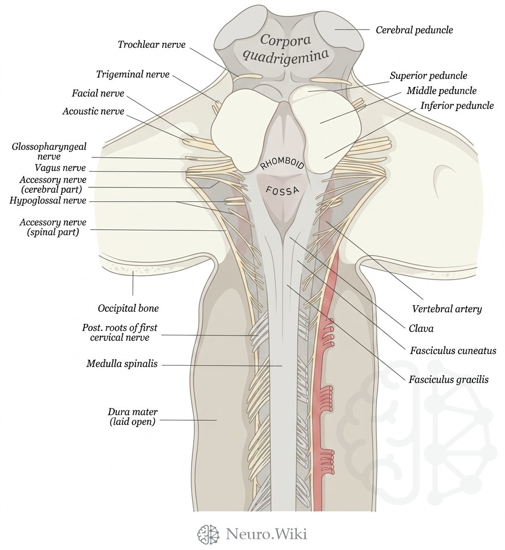
Posterior view of upper spinal cord, hindbrain and midbrain.
The medulla oblongata is the caudal segment of the brainstem, continuous above with the pons and below with the spinal cord at the foramen magnum. Its dorsal surface forms the lower rhomboid fossa, floor of the fourth ventricle, while the closed caudal portion carries paired longitudinal columns — the fasciculus gracilis medially and the fasciculus cuneatus laterally — each expanding into the clava and cuneate tubercle over their dorsal column nuclei. The inferior cerebellar peduncle ascends along its posterolateral aspect. Rootlets of the glossopharyngeal, vagus, and cranial accessory nerves emerge from the postolivary sulcus, with hypoglossal rootlets exiting more ventrally.
Structures labelled on this plate
- Trochlear nerve
- Cerebral peduncle
- Corpora quadrigemina
- Trigeminal nerve
- Superior peduncle
- Facial nerve
- Middle peduncle
- Acoustic nerve
- Inferior peduncle
- Glossopharyngeal nerve
- Vagus nerve
- Accessory nerve (cerebral part)
- Hypoglossal nerve
- Rhomboid
- Fossa
- Accessory nerve (spinal part)
- Occipital bone
- Vertebral artery
- Clava
- Posterior roots of first cervical nerve
- Fasciculus cuneatus
- Medulla spinalis
- Fasciculus gracilis
- Dura mater (laid open)
Source: After Gray's Anatomy (1918). Public domain (Gray's Anatomy, 1918), modernized rendering.
Rendering © 2026 Neuro.Wiki — recolouring, relabelling and three-dimensional modelling. The underlying historical figure is public domain and remains so. Free to view and to cite with a link; not licensed for redistribution, dataset compilation, or AI model training.