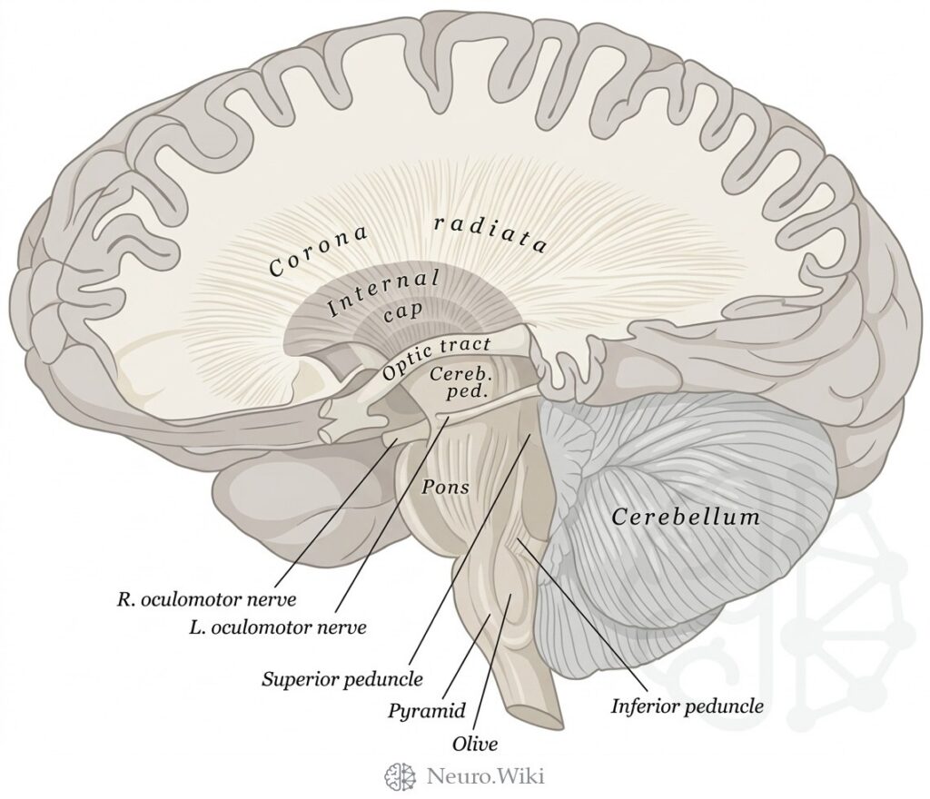
Lateral dissection showing corticospinal fibers
About this plate
Lateral dissection showing corticospinal fibers.
A midsagittal section of the brain exposes the medial surface of one cerebral hemisphere together with the brainstem, cerebellum and upper spinal cord. The corpus callosum arches above the diencephalon, its rostrum, genu, body and splenium framing the sulcus of the corpus callosum and, below, the subcallosal gyrus and paired parolfactory sulci. The lamina terminalis descends from the anterior commissure to the optic chiasma and infundibulum, while the hypothalamic sulcus separates thalamus from hypothalamus. Posteriorly lie the pineal body, posterior commissure and lamina quadrigemina. The midbrain continues into pons and medulla oblongata, forming the floor of the fourth ventricle beneath the cerebellum.
Structures labelled on this plate
- Corona radiata
- Internal cap
- Optic tract
- Cereb. ped
- Pons
- Cerebellum
- R. oculomotor nerve
- L. oculomotor nerve
- Superior peduncle
- Pyramid
- Olive
- Inferior peduncle
Source: After Gray's Anatomy (1918). Public domain (Gray's Anatomy, 1918), modernized rendering.