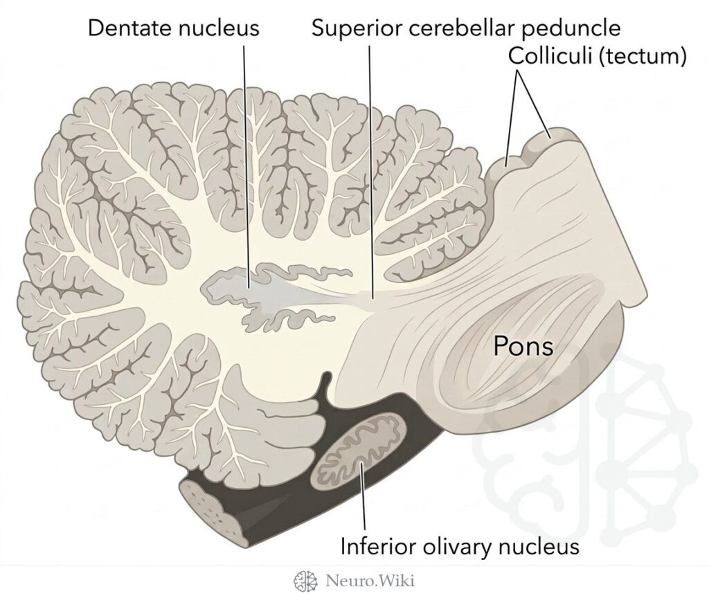
Sagittal section through right cerebellar hemisphere and inferior olive
About this plate
Sagittal section through right cerebellar hemisphere and inferior olive.
The cerebellum occupies the posterior cranial fossa, dorsal to the pons and medulla and separated from the occipital lobes by the tentorium cerebelli. In sagittal profile its surface is folded into narrow, transversely oriented folia grouped into lobules, with the arbor vitae of white matter branching into each folium. Deep within each hemisphere lies the dentate nucleus, a crumpled sheet of grey matter whose efferents emerge as the superior cerebellar peduncle, ascending toward the midbrain tegmentum beneath the paired colliculi of the tectum. Anteriorly the pons bulges forward, while the inferior olivary nucleus forms a bulge on the ventrolateral medulla.
Structures labelled on this plate
- Dentate nucleus
- Superior cerebellar peduncle
- Colliculi (tectum)
- Pons
- Inferior olivary nucleus
Source: After Gray's Anatomy (1918). Public domain (Gray's Anatomy, 1918), modernized rendering.
Rendering © 2026 Neuro.Wiki — recolouring, relabelling and three-dimensional modelling. The underlying historical figure is public domain and remains so. Free to view and to cite with a link; not licensed for redistribution, dataset compilation, or AI model training.